DAY 5
Right when we came into lab we took out our environmental
samples to observe the bacterial growth. There was a significant amount of
growth on the plate. It looks like Grace doesn’t brush as well as we thought!

Simple Stain:
First we learned how to prepare a simple stain. We prepared
a fixed smear (using our bacteria from day 1/2) and once it was dry we added
crystal violet stain. After that, we rinsed the slide and then blotted it with
bibulous paper. Using oil immersion, we were able to analyze the bacteria under
the microscope. Our bacteria appeared to be rod-shaped.


Gram Stain:
Next we learned about gram staining. We put a fixed smear of
bacteria on a slide over a staining tray on a sink rack. Then we put crystal
violet smear on it for 20 seconds, and then rinsed off the excess. We covered
the smear with gram iodine for one minute. Then we rinsed the slide to remove
the excess. We decolorized it with 95% ethanol and held it at an angle and
added decolorizing reagent drop by drop until color stops running. We then
rinsed the slide to remove the decolorizing agent, and covered it with safranin
for one minute. We rinsed the slide to remove the excess and blotted it with
bibulous paper and examined it under the microscope using oil immersion. After
looking at the bacteria under the microscope we determined our specimen was
gram-negative.


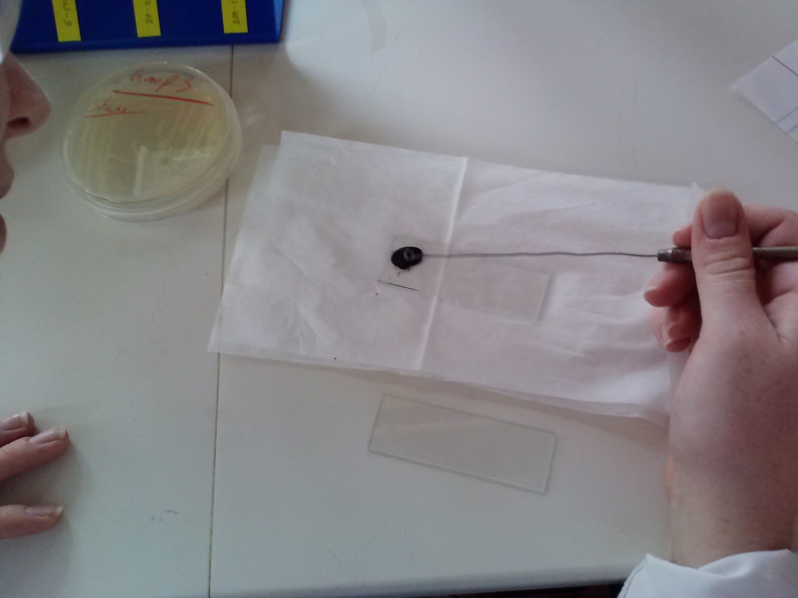
Gram negative staining:
The next thing we did was gram negative stating. We put a
drop of nigrosin on the slide first, and then got bacteria from our spread and
mixed it together. We took another slide and slid it across so it spread onto
the entire slide. We then let it dry and examined it under the microscope.


In conclusion today, we stained bacteria with three different techniques: Simple Staining, Gram Staining, and Gram Negative Staining. Here are our three different slides!

No comments:
Post a Comment